BLOG CONSOPHARMA

Cervical Radiculopathy Causes, Symptoms, and Treatment

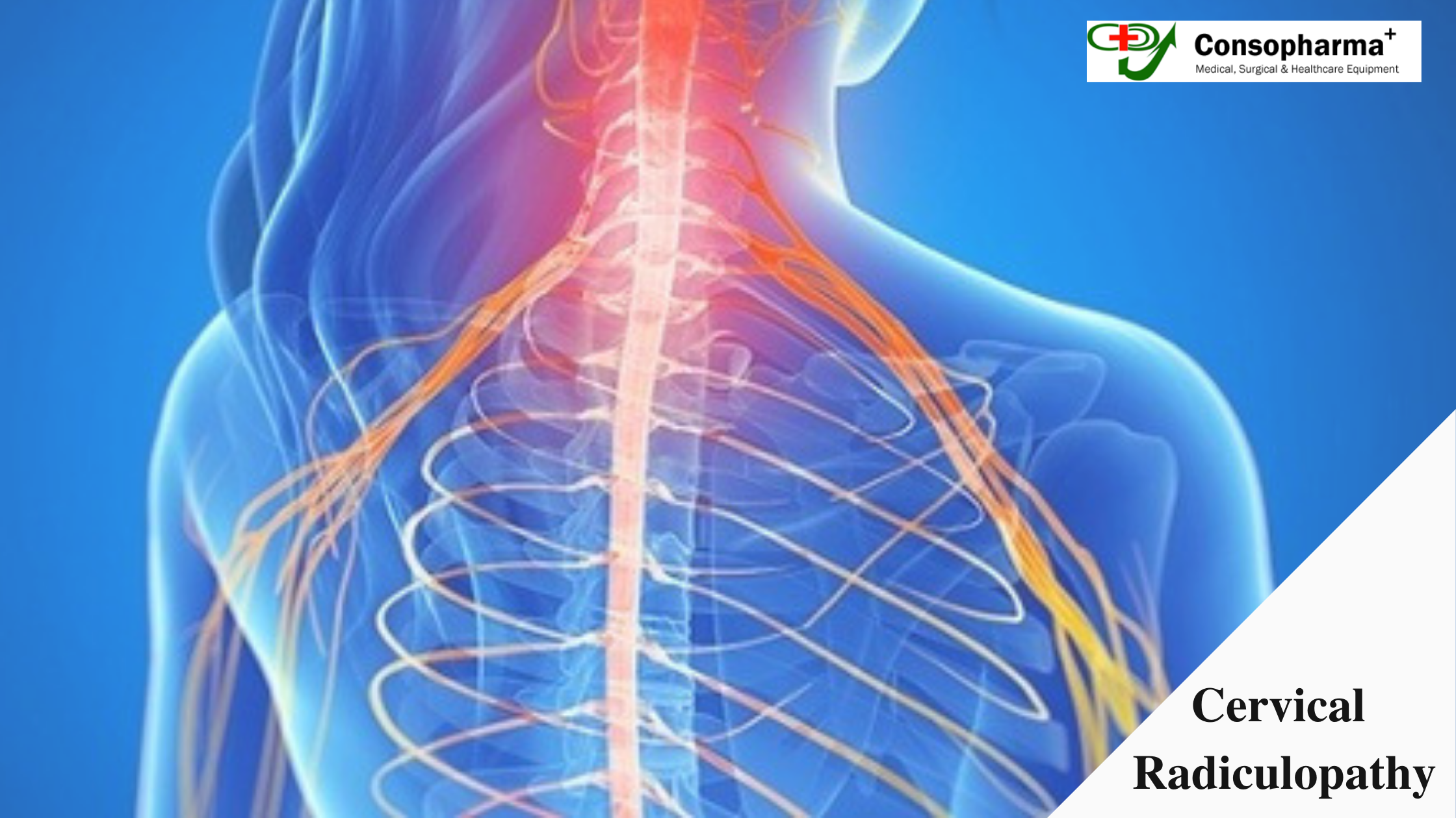
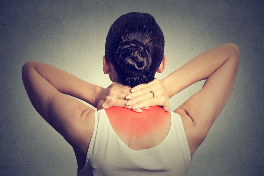
Cervical Radiculopathy is a condition in which the nerve in the neck which is called cervical spine is damaged or causing pain. The doctor generally calls this condition Cervical Radiculopathy. The nerves in the spine leave the spinal section through openings during the bones of the spine (vertebrae) from the right and left sides. These nerves are exciting from the spinal canal. They are numbered from 1 to 8. This is based on the same vertebrae numbering. It starts from the bottom of the skull. The condition Radiculopathy can cause diverse agony, shivering and numbness along the shoulder. Also, it causes numbness along the arms and hands. It depends on the nerve which is damaged.

If you are suffering from Radiculopathy then you should know some of the symptoms and the treatments. You have to visit a doctor to get it treated and to manage the pain which is caused by Radiculopathy.

Cervical Radiculopathy Causes

Cervical Radiculopathy is caused generally to older people because of the natural changes like age. In younger people, it is generally caused because of a sudden injury.

Main Causes of Radiculopathy are:-
  • 1. Cervical Degenerative Disc DiseaseIn this condition, the celebrated disk loses the natural liquid it has; in the result, it shrinks or develops cracks at the exterior layer of the disc. This condition can start with an accident or with everyday work.
  • 2. Cervical Herniated DiscIt is also known as a disc bulge. It occurs when the gel-like focus crushes out of a tear or a split in the external ring of the disc. It can cause Radiculopathy due to the pressure on the nerve root. Generally, the people suffering from this disease have numbness, weakness in neck, shoulders and arms.
  • 3. Cervical Spinal StenosisIn this condition, the spinal canal becomes narrow and that squeezes the spinal cord. This disease causes some of the symptoms like numbness in the hand or in the arm, neck pain, and problems with walking. Also, there is a problem with the balance during walking.

Some of the symptoms related to Radiculopathy are:
  • 1. C5 Radiculopathy
  • 2. C6 Radiculopathy
  • 3. C7 Radiculopathy
  • 4. C8 Radiculopathy

Treatment For Cervical Radiculopathy

Non-surgical changes
  • 1. Change in lifestyleSome of the minor symptoms related to Radiculopathy can be cured by its own. Sports activities should be stopped. Ice packs should be applied to the neck and may also feel relief.
  • 2. Physical TherapyPhysical therapists can help to cure this disease. If a patient does stretching and exercises it will help to reduce the effect of Radiculopathy.

Surgical changes
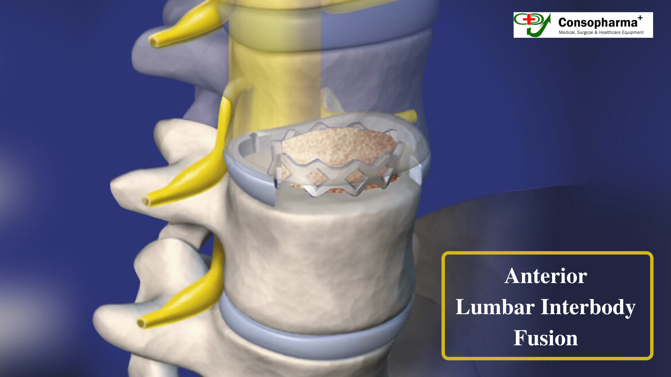
  • 1. Cervical Discectomy and FusionThis is a surgery which can relieve radiculopathy. In this, the unhealthy dick is removed and in place of it, a plastic implant filled with bone will be planted.
  • 2. Artificial Disk ReplacementIn this treatment, the damaged cervical disc is replaced by an artificial disc. It will reduce pain and may help the patient to do all the everyday work. So choose and buy the implants from the best Cervical Implants Supplier.

CONTACT US

All Your Queries & Suggestions Are Most Welcome. Feel Free to Contact Us Anytime.

Please call and whatsapp +919350504074 if urgent

your personal case manager will ensure thate you receive the best possible care

BLOG CONSOPHARMA

Anterior Lumbar Interbody Fusion – The Best Treatment for Disc Problems

In Anterior lumbar interbody fusion, a bone graft is placed in the free space through an incision through the belly. The disc is removed through the incision, and graft is used to substitute for the height. Upon healing, this becomes a consolidated piece. This method is used to correct all disc related problems to realign bones.

Advantages
  • As this method uses more space, the support it guarantees is also large with increased healing power.
  • The spine is reached through the incision in the belly and hence the back muscles are not disturbed, causing lesser pain. This also allows the surgeon to work without meddling with the spinal nerves.

What happens during the surgery?

The surgery which is performed together by a neurosurgeon or an orthopedic surgeon, along with the help of a vascular surgeon. The surgery which takes close to 1 to 2 hours is comprised of the following steps.

  • 1. Prepare the patient and give anaesthesia. The surgery is conducted through an incision in the belly and hence belly is cleansed and prepared.
  • 2. Using an X-ray fluoroscope or image guidance, an incision of 2-3 inches at the disc level is made in the belly.
  • 3. Now, the vascular surgeon makes path to locate the damaged disc by clearing the path between the abdominal cavity and the retroperitoneal space. The intestines are shifted temporarily to the right side of the belly . Similarly, the veins and arteries are also moved to a side.
  • 4. The spine surgeon comes to rescue here for removing the damaged disc. The damaged disc is opened inserting a distractor instrument and the fusion bed is prepared using bone shavers.
  • 5. Now, the trial spacer is placed in the empty disc space after measuring the open disc space and selecting a spacer size. The surgeon now makes sure that everything like placement, height, wedge angle is accurate and the nerves can decompress, using an x-ray. After this, the bone graft material is made ready for fusion. This paste is mortar like but contains bone growing proteins packed into permanent bioplastic spacer cage.
  • 6. Once again with the help of x-ray fluoroscopy, this spacer graft is placed in the empty disc space. The two bones are pushed apart so that the normal disc height is restored.
  • 7. The next step is to insert the plate and screws so that the spacer graft is secured tightly in place either screws or with a metal plate which can be screwed into the front portion of the vertebrae. The titanium bone plate can be used which guarantees stability. The right choice has to be made while deciding the spine instruments suppliers or bone locking screw suppliers as even slight carelessness could cause complications.
  • 8. The surgery is over, and the peritoneum restores back to it’s position. The abdominal muscles are closed with sutures and the incision with skin glue.

Causing minimal trauma to spine and the organs and tissues surrounding it, the ALIF promises a speedy recovery.

CONTACT US

All Your Queries & Suggestions Are Most Welcome. Feel Free to Contact Us Anytime.

Please call and whatsapp +919350504074 if urgent

your personal case manager will ensure thate you receive the best possible care

BLOG CONSOPHARMA

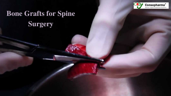
Different types of bone grafts in Spine surgery

Bone grafting is a medical or specifically a surgical procedure that is used to fix deformities or disparities caused to bones and joints. Some severe accidents and high impact injuries leave victims’ bones damaged severely so that a bone grafting or a tissue transplant is required to take back the victim to normal life. Bone grafting is also used to help a bone grow near an orthopaedic implant device. Bone structure deformities caused by birth or by accidents where a specific bone or a group of bones is completely absent are cured by bone grafting procedures, and this becomes possible with the help surgical equipment suppliers.

Why is bone grafting practised?

Bone grafting is done for a number of reasons, from curing injuries to diseases. There are several reasons for the need for bone grafting

  • In case of a spinal injury, commonly a joint gets deceased and thus to help the two bones across the joint heal, a fusion process is carried out.
  • In some severe accidents, patients lose some specific bone. Loss of a bone may even be caused by a disease. In such a scenario, bone grafting is used to regenerate that specific bone with the help of spine implant instruments.
  • Joint replacements or spinal fusions have surgical orthopaedic implants supporting the bine structure. Bone grafting is used around these implants to grow bones.

Bone grafts in spine surgery

Spinal fusion is generally performed on the neck or back. It is done in order to get the patient relieved of the pain that is caused by two struggling bones. So, the bones are fused so that they can heal into a single bone. For this purpose, an additional bone known as the bone graft is used.

Autograft bone graft

This kind of spinal fusion involves the patient’s own bone being the bone graft. The common donor area is the iliac crest located in the patient’s pelvis.

Allograft bone graft

In this kind of bone graft, bone is obtained from cadavers for use in the spine fusion. Different techniques and measures are employed to sterilize the donor’s bone, and they are also available in different shapes and sizes.

Local bone graft

To release the pressure on some nerve roots, spinal bones are removed. During spinal fusions, these removed bones are used as a bone graft.

Synthetic bone

These bones are made with calcium materials. They are used as substitutes to autograft bone implants. Spine implant suppliers often provide them with other spine implants.

Now, that you are well aware regarding the different types of bone grafts in spine surgery making a final decision will be that much easier for you.

CONTACT US

All Your Queries & Suggestions Are Most Welcome. Feel Free to Contact Us Anytime.

Please call and whatsapp +919350504074 if urgent

your personal case manager will ensure thate you receive the best possible care

BLOG CONSOPHARMA

Knee Arthroscopy- A surgical procedure for knee joint

Knee arthroscopy is a medical procedure that is carried out on patients suffering from knee joint problems. Unlike the major open surgeries, this method is proceeded by the help of exceedingly small incisions made on the required body part. A tiny camera, which is also known as an arthroscope, is inserted in the knee. This helps the surgeon to examine the inside of the knee and investigate the complexity of the problem. The images of the knee obtained by the arthroscope helps the surgeon to make the necessary corrections in the bones and the tissues.

Anatomy behind the procedure

The knee joint in the human body is one of the biggest joints in the body and is in fact regarded as one of the most complex joints. Orthopaedic implants and instruments have by far helped the surgeons to revert back the deformities caused to the knee joint. The lower end of the femur and upper end of the tibia and patella makes up the knee. Since three major bones are involved with the knee joint, the surgical operations become a bit tedious.

  • Articular cartilage – The ends of the femur and tibia are covered with this cartilage.
  • Synovium – A thin lining called synovium surrounds the knee joint.
  • Meniscus – The meniscal cartilage acts like the shock absorbers.
  • Ligaments – Ligaments are known as bone connectors.

Surgical procedure

Surgical draping is covered all over the patient’s leg so as to expose the prepared incision site. The skin of the knee is cleaned in order to prevent any surgical infections. Anaesthesia dose will be given to the patient so that the patient does not feel any pain in the surgical portion. The surgeon starts small incisions or cuts in the knee, and sterile saltwater will then be pumped into the knee to expand the surgical portion for clarity. The arthroscope will be entered into the knee, and the surgeon will look at the affected portion on the monitor.

When the surgeon locates the problems in the knee, he/she will make certain cuts on the required tissue to correct the deformity and will drain out the saline water out. Surgical implant suppliers such as orthopaedic bone screws distributors have made it possible to carry out major knee surgeries that require utmost precision.

Surgical equipment suppliers provide the necessary apparatus for the recovery period of the surgery. Though the operation is not must invasive, and knee dressing materials help in fast recovery.

CONTACT US

All Your Queries & Suggestions Are Most Welcome. Feel Free to Contact Us Anytime.

Please call and whatsapp +919350504074 if urgent

your personal case manager will ensure thate you receive the best possible care

BLOG CONSOPHARMA

Anterior Cervical Discectomy & Fusion-Surgery For Chronic Neck Pain

Anterior cervical discectomy and fusion (ACDF) is a surgery done to remove a degenerative or herniated disc from the neck. It is a surgery that helps to reduce or eliminate chronic pain in the neck and back due to problems with the discs. The surgery is anterior because the surgeon accesses the disc through the front of the neck instead of the back. There are many spine implant suppliers who supply various instruments for surgery, and with these instruments, the procedure can be carried out smoothly.

The Procedure

The procedure is simple, and the surgeons use instruments and devices that are supplied from the bone locking screw suppliers. The steps of the procedure include: –

  • The surgeon makes a small incision at the front of the neck and then moves aside the neck muscles and the airway, exposing the patient’s disc and vertebrae.
  • After this, they use a special X-ray and locates the affected vertebrae and discs.
  • Depending on the condition of the patient and with the help of the surgical tools, the surgeon then removes one or more discs.
  • Also, any bone spurs pressing on the nerve roots and surrounding the damaged discs are removed as well.
  • Bone graft material, which is derived from a patient, a cadaver, or a man made product, is inserted into the empty disc space. These bone grafts may be fixed in place with metal plates and screws.
  • After the process, the incision is closed, and the patient is placed in the post-operative recovery area.
What to expect in ACDF?
  • It is usually an outpatient procedure, and so the patient can return home on the day of their surgery. This operation takes place under general anesthesia and so the person will be asleep during the surgery.
  • After the surgery, most of the patients will experience some pain, but it can be managed by taking pain killers. However, the recovery can take up several weeks, and some people might have to wear a collar in order to stabilize their neck.
  • Also, most of the people might have to meet their surgeon 4 to 6 weeks after their surgery for an assessment. And they might be asked to avoid strenuous activity until recommended by the surgeon.

ACDF carries some risks, but it is relatively uncomplicated, and so before you opt for a surgery consult with the surgeon and the cervical implants supplier for good quality implants.

CONTACT US

All Your Queries & Suggestions Are Most Welcome. Feel Free to Contact Us Anytime.

Please call and whatsapp +919350504074 if urgent

your personal case manager will ensure thate you receive the best possible care