Anterior Lumbar Interbody Fusion – The Best Treatment for Disc Problems
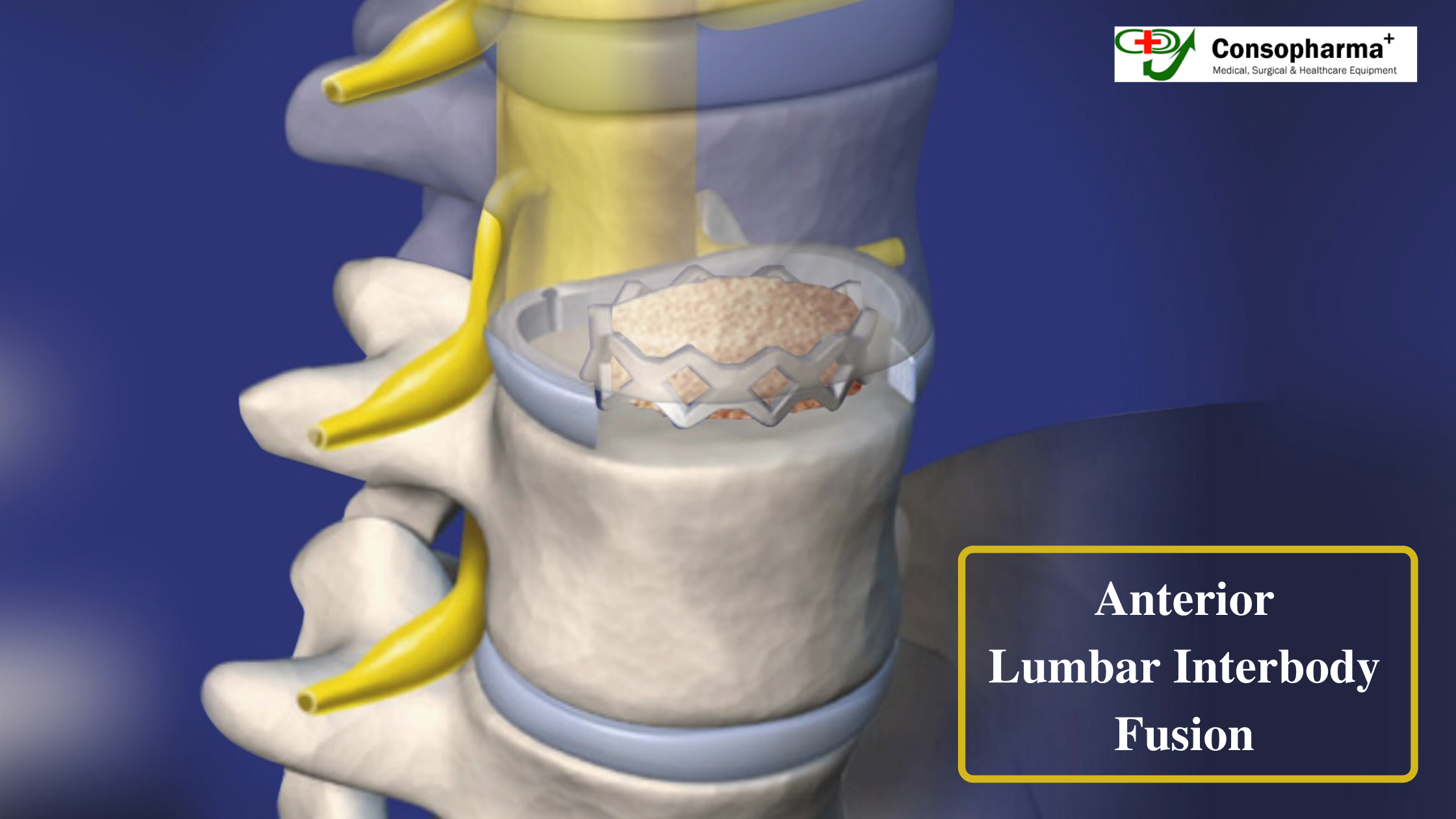
In Anterior lumbar interbody fusion, a bone graft is placed in the free space through an incision through the belly. The disc is removed through the incision, and graft is used to substitute for the height. Upon healing, this becomes a consolidated piece. This method is used to correct all disc related problems to realign bones.
Advantages
- As this method uses more space, the support it guarantees is also large with increased healing power.
- The spine is reached through the incision in the belly and hence the back muscles are not disturbed, causing lesser pain. This also allows the surgeon to work without meddling with the spinal nerves.
What happens during the surgery?
The surgery which is performed together by a neurosurgeon or an orthopedic surgeon, along with the help of a vascular surgeon. The surgery which takes close to 1 to 2 hours is comprised of the following steps.
- 1. Prepare the patient and give anaesthesia. The surgery is conducted through an incision in the belly and hence belly is cleansed and prepared.
- 2. Using an X-ray fluoroscope or image guidance, an incision of 2-3 inches at the disc level is made in the belly.
- 3. Now, the vascular surgeon makes path to locate the damaged disc by clearing the path between the abdominal cavity and the retroperitoneal space. The intestines are shifted temporarily to the right side of the belly . Similarly, the veins and arteries are also moved to a side.
- 4. The spine surgeon comes to rescue here for removing the damaged disc. The damaged disc is opened inserting a distractor instrument and the fusion bed is prepared using bone shavers.
- 5. Now, the trial spacer is placed in the empty disc space after measuring the open disc space and selecting a spacer size. The surgeon now makes sure that everything like placement, height, wedge angle is accurate and the nerves can decompress, using an x-ray. After this, the bone graft material is made ready for fusion. This paste is mortar like but contains bone growing proteins packed into permanent bioplastic spacer cage.
- 6. Once again with the help of x-ray fluoroscopy, this spacer graft is placed in the empty disc space. The two bones are pushed apart so that the normal disc height is restored.
- 7. The next step is to insert the plate and screws so that the spacer graft is secured tightly in place either screws or with a metal plate which can be screwed into the front portion of the vertebrae. The titanium bone plate can be used which guarantees stability. The right choice has to be made while deciding the spine instruments suppliers or bone locking screw suppliers as even slight carelessness could cause complications.
- 8. The surgery is over, and the peritoneum restores back to it’s position. The abdominal muscles are closed with sutures and the incision with skin glue.
Causing minimal trauma to spine and the organs and tissues surrounding it, the ALIF promises a speedy recovery.
Recent Posts
-
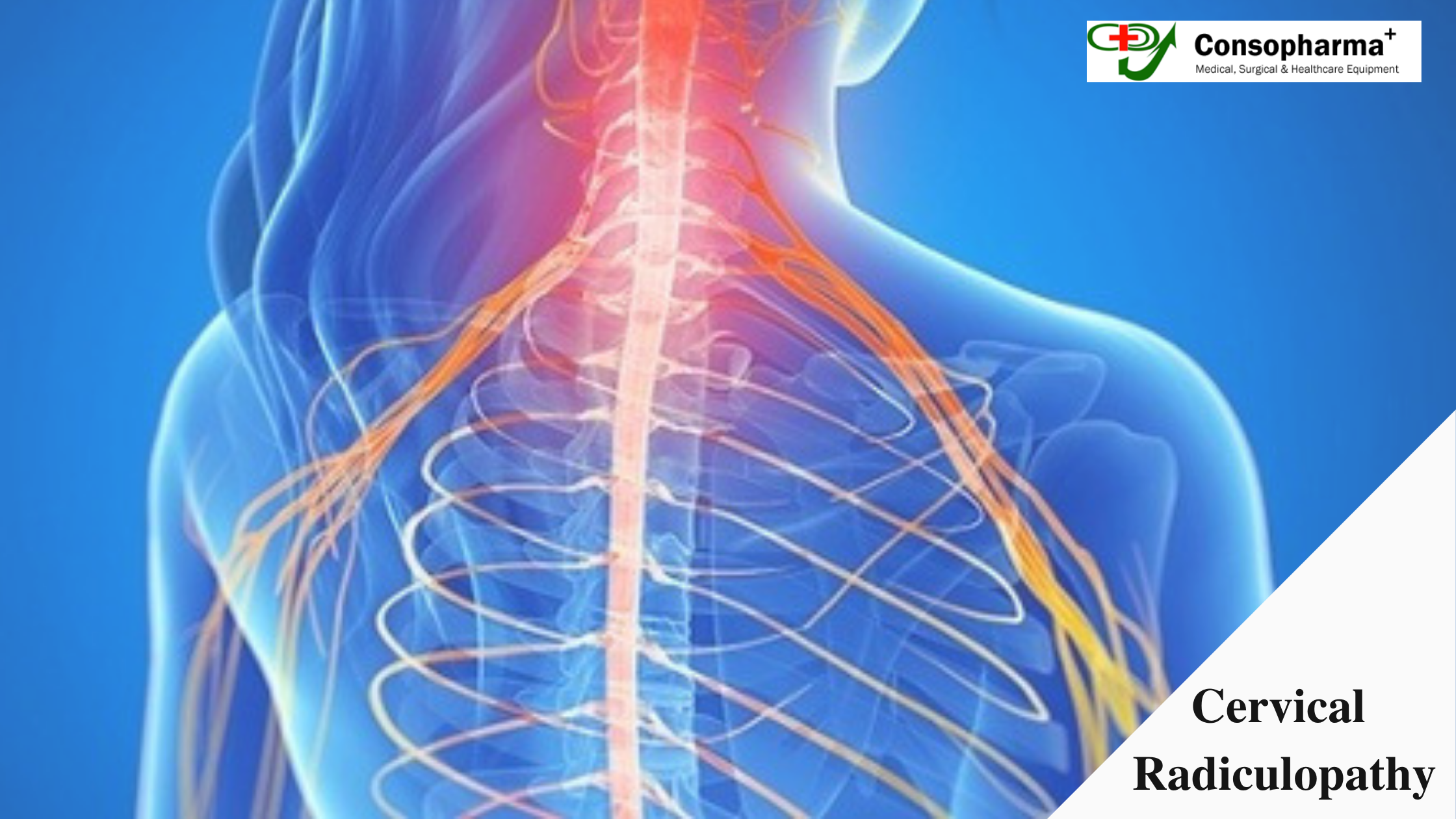 Cervical Radiculopathy Causes, Symptoms, and Treatment July, 13 2020
 -
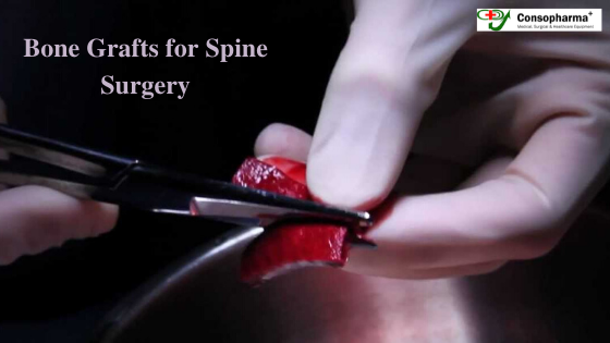 Different types of bone grafts in Spine surgery June, 6 2020